The Portable Ultrasound Scanners project—running from 2013 to 2018—has received DKK 75 million (EUR 10 million) in funding from Innovation Fund Denmark out of a total budget of DKK 149 million (EUR 20 million).
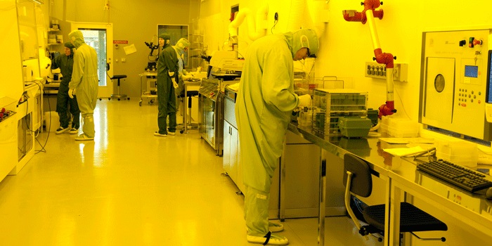
From the outside, it looks like a space station engulfed in yellow light, where humanoid figures walk around purposefully between transparent cabinets with robotic arms juggling CD-like discs and man-sized microscopes connected to computers and monitors. It is not easy to tell whether they are men or women, because they are all covered in boiler suits, large bulky overshoes, glasses, and caps pulled down over their foreheads.
If you want to enter the clean room, there is no way around the unsexy and unbearably warm outfit, which also includes disposable gloves pulled over the suit sleeves. Packed in sealed bags, the outfit may only be opened inside the dressing room or the sluice, as it is called. And you should preferably not touch it on the outside—no easy task for a first-time visitor.
Here, a day’s work may be ruined by a grain of dust. That is why you have to go through the sluice and the cumbersome change of clothes, and why all air is blown into the room through efficient filters in the ceiling and is sucked out through the perforated floor. The air which usually surrounds us contains 1 million particles per cubic meter. In the clean room, this figure is reduced to 100. It is, in other words, not completely dust-free, so running is strictly forbidden, as it will stir up the particles.
Shoes, phones, dictaphones, and pens must be left outside. All communication with the outside world takes place via landline phones attached to the walls, and any notes must be made on special non-fluffy paper using dusted pens, which are available in the sluice.
And now you are ready for a tour of DTU Danchip. I’m going to see how the transducer—the vital part of an ultrasound scanner—is made. The transducer converts electrical signals to sound waves and vice versa, when the sound returns as an echo from the interior of the body. The transducer must thus be able to emit and absorb sound and be made of a material which can be made to oscillate by an electrical pulse.
You would normally use a so-called piezoelectric crystal. It works fine, but the technology is limited by the fact that the distance between the individual transducer elements cannot be reduced to much less than 30 micrometres, and that crystals contain lead.
"Many of the large ultrasound companies have tried to make these silicon transducers, but have given up. However, we’re well under way and have already made the first fully functional probes."
Professor Erik Vilain Thomasen, DTU Nanotech
Therefore, experiments are made to find an alternative, which is both made of the harmless material silicon and which can be made extremely small. This means that the critical dimensions may routinely be kept at around a micrometre.
I therefore don’t stand a chance to see what it’s actually all about. What I can see is something similar to CD discs. These so-called wafers, slices cut from silicon rods, will be placed in one of the transparent cabinets. A robot arm grabs a wafer and places it in a holder where it is held firmly in place by vacuum. And while the wafer is turned, various fluids are distributed on it.
First a metal layer intended to serve as base contact. Then an insulating polymer layer, where UV light in a so-called photolithography process forms a pattern of insulators, a kind of ‘bricks’ interspersed with air. The photosensitive polymer cannot withstand white light, which is why the entire clean room is swathed in yellow.
The top layer is an ultra-thin silicon wafer, to which is added a top electrode. When you later apply AC voltage over the electrodes, the silicon wafer will start vibrating and thus emit the sound, which this is all about.
When the layers are in place, the wafer is carried to the processing chamber where it is baked at 110°C. It is then cooled to room temperature, and a developer is applied to it. Finally, the developer is rinsed off with water, and the wafer is now ready to be checked under the microscope.
Devil in the detail
Each wafer may have several transducer elements, which consist of a pattern of tiny ‘drums’, each having their own contact. They are also known as CMUTs, an abbreviation for Capacitive Micromachined Ultrasonic Transducers.
“The smaller the CMUT cell is, the higher the frequency at which the final probe must be operated. The processes in the clean room give us a lot of freedom to determine the size and design of the CMUT cells, and this technology is therefore well suited making compact ultrasound transducers and high-frequency probes,” says PhD student Andreas Havreland from DTU Nanotech, who is my guide in the clean room.
It is therefore a question of generating the exact frequency suitable for a particular medical purpose. The higher the frequency, the better the resolution of the image, but the more sound you also lose in the process. High frequencies penetrate less far into the body than low frequencies, which most people have probably experienced, when their neighbour throws a party, and the bass travels through walls easier than other frequencies.
DTU researchers have designed a structure that has proven to work in both a 2D probe where the transducers are only placed in one direction, and in a 3D probe where they are located so that the sound is emitted in two directions. But there is still a lot of work to do in optimizing the structure, so that the sound pressure is high enough to penetrate deep into the body of also heavy people.
“Many of the large ultrasound companies have tried to make these silicon transducers, but have given up. However, we’re well under way and have already made the first fully functional probes, and I’m certain that we—within the project time frame—will be making even better probes, which can emit higher pressure waves,” says Professor Erik Vilain Thomsen, DTU Nanotech.
The daily production output is inspected under the microscope where the fine patterns can be clearly seen when magnified 10 times. Yes, the patterns have the desired size, and the electrodes are placed exactly where they should be. So these transducer elements are ready to be sliced and placed in a probe.
And today’s visitor to the clean room is ready to return to the dusty, white world.